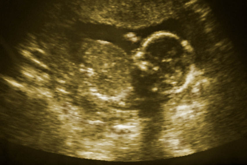
Ultrasound Scans
Ultrasound scans are an excellent diagnostic tool used in the field of medicine. This article focuses on its uses, types, procedure, care to be taken before, during and after a scan, as well as the advantages and disadvantages of ultrasound scans.
DEFINITION
Ultrasound, sonography, or ultrasound scanning, is a non-invasive diagnostic test which uses sound waves to produce images of the important body parts.
USES
Ultrasound scans may be used for the following:
- Abdominal scans – To investigate abdominal-related symptoms and examine structures in the abdomen.
- Pelvic scans – To identify the cause of pelvic pain or conditions associated with the female reproductive system.
- Pregnancy scans – To determine the due date, verify fetal abnormalities, as well as monitor the growth and progress of the fetus.
- Other uses – To be used for musculoskeletal scans, breast scans (to scan for cancers), or Doppler ultrasound (which detects the speed and blood flow in the body’s arteries and veins).
TYPES OF ULTRASOUND SCANS
There are three primary types of ultrasound scans.
- External ultrasound scan – in which the probe is externally used. It is used to examine internal organs such as the heart, pelvic and abdominal organs and the fetus’s health.
- Internal or transvaginal ultrasound scan – in this a thin probe is inserted inside the body via the vagina or anus. This scan type can be used to inspect the ovaries, uterus or prostate gland.
- Endoscopic ultrasound scan – in this an endoscope (long and flexible tube) is attached to the probe, enabling it to move further in the body. This enables improved viewing of internal organs such as the stomach or oesophagus (food pipe).
PROCEDURE
Most ultrasound scans are performed with the patient lying on their back on an examination table. A gel is then applied to the area being examined, which helps in proper contact of the device with the skin for improved imaging. The radiologist or sonographer then slowly moves the transducer across the skin and analyzes images which form on the computer screen. In certain cases, for improved imaging, the transducer is inserted in the body. Moreover, to view the heart-related images, the transducer may be inserted in the oesophagus (called transesophageal echocardiogram). If the transducer is rectally inserted, the man’s prostate can be viewed (called transrectal ultrasound). If the transducer is inserted in the vagina, a woman’s ovaries and uterus can be visualized (called transvaginal ultrasound).
PREPARING FOR THE ULTRASOUND SCAN
To enhance the visualization of specific organs, specific preparations may be necessary.
- Drink a considerable amount of water and avoid going to the toilet until after the scan – This is usually performed when conducting pregnancy or pelvic region scans.
- Staying nil by mouth for several hours before the scan – may be required for scanning organs of the digestive system.
- You may be asked to change to a hospital gown.
- An anesthetic agent may be given for transesophageal scans. This is usually sprayed at the back of your mouth to numb the area and allow an easy passage of the scope.
- Often, a contrast agent (a harmless substance injected in the vein) may be injected to view additional detailed images.
IMMEDIATELY AFTER THE ULTRASOUND
Usually, an ultrasound scan lasts for 20–40 min depending on the body part being examined. After the procedure, you will be given paper towels to wipe off the gel. You can then change back to your regular clothes. The results of the ultrasound scan will then have to be discussed with your doctor during the same or a follow-up appointment.
HOME CARE AFTER THE ULTRASOUND SCAN
The procedure for ultrasound is painless; you can resume your normal activities at home after the scan.
ADVANTAGES AND DISADVANTAGES OF ULTRASOUND SCANS
- Ultrasound scans have an excellent safety record.
- It does not require ionizing radiation and therefore has lesser risks compared to other imaging techniques such as X-rays.
- It is safe when cautiously used.
- To some extent, ultrasound waves can heat body tissues, resulting in small gas-filled pockets in tissues or body fluids. The long-standing effects are still unidentified; therefore, it is important to judiciously use ultrasound scans during pregnancy.
Ultrasound scans have been an effective diagnostic investigation because they have been mostly non-invasive and use non-ionizing radiation. Moreover, they have been a boon to the medical fraternity.
References
- Cancer Quest. Ultrasound Imaging [Internet] [Updated Jan, 2020]. Available at: https://www.cancerquest.org/patients/detection-and-diagnosis/ultrasound. Accessed on May 11, 2021.
- Better Health. Ultrasound scan [Internet] [Updated Feb 6, 2019]. Available at: https://www.betterhealth.vic.gov.au/health/ConditionsAndTreatments/ultrasound-scan. Accessed on May 11, 2021.
- NHS. Ultrasound scan [Internet] [Updated May 25, 2018]. Available at: https://www.nhs.uk/conditions/ultrasound-scan/. Accessed on May 11, 2021.
- FDA. Ultrasound Imaging [Internet] [Updated Sep 28, 2020]. Available at: https://www.fda.gov/radiation-emitting-products/medical-imaging/ultrasound-imaging. Accessed on May 11, 2021.

