Ureteropelvic Junction Obstruction in Newborns: Symptoms, Causes and Treatment Recognizing Symptoms of Ureteropelvic Junction Obstruction in Newborns Our effortlessly perfect human body may at times develop some congenital conditions. Recognizing these and treating them on time can be life-saving.
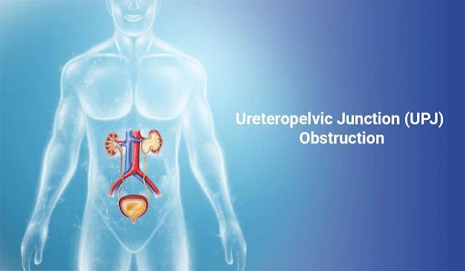
In a healthy scenario, the urine is formed in the kidneys, from where it flows to the ureter (a tube carrying urine) and the urinary bladder (the organ that stores urine).
Ureteropelvic junction (UPJ) Obstruction is a condition where the flow of urine from the renal pelvis (a funnel-shaped portion of the kidney) to the ureter is partially or completely blocked. This blockage can lead to the accumulation of urine in the kidneys. If left untreated it can lead to kidney damage, making early recognition and intervention essential, particularly in newborns.
What causes UPJ Obstruction?
UPJ obstruction can be caused by an abnormality in the muscle or tissue at the junction, external compression, or, less commonly, a blood vessel crossing over the junction. It could be genetically linked and is congenital in most cases. However, no specific gene alterations are known for this condition. The obstructions present at birth can be:
Abnormal muscle cell arrangement at the ureter: This prevents the urine from being pushed down
Narrow ureter: Obstructing urine flow
Abnormality in connection of ureter to the pelvis: Ureter may connect to the pelvis at a junction that obstructs normal flow.
Unusual folds in the ureter: These may act as valves and prevent normal flow of urine.
Presence of extra blood vessels: In some cases, the child may have a crossing vessel, which means an extra artery. This can cause kinking or blockage of the ureter.
How will a parent know if the child has UPJ obstruction?
A parent may often worry if their newborn is in good health. Identifying any underlying condition early on is paramount to make sure any underlying condition is identified at the earliest at the right cure is administered. UPJ obstruction, in case of congenital and developmental conditions, can be mild, moderate, or severe and can present with varying symptoms. Diagnosis before the child is born: Since the renal system develops in the fetus, a UPJ obstruction if causing major obstruction can present in prenatal ultrasound done by a fetal medicine expert. In case the doctor finds signs of hydronephrosis (swelling of the kidney) in the child then UPJ obstruction could be suspected. No treatment is done at this stage, and the baby is vigilantly monitored in subsequent fetal ultrasounds and post-birth to decide if treatment is needed.
Diagnosis in a newborn post-birth: Mild cases of UPJ obstruction may not present with any symptoms, and if it is detected in prenatal or postnatal ultrasound such a condition may resolve on its own in many cases. Moderate or severe obstruction may present with specific symptoms, which should be identified early on to ensure that the right treatment is done and the damage to kidneys is prevented.
Postnatal Symptoms include:
Lump in the abdomen: A swollen kidney might be palpable.
Frequent urinary tract infections (UTIs): Infections may occur due to urine stagnation.
Vomiting or poor feeding: Signs of discomfort or systemic impact of the condition.
Blood in the urine (hematuria): This may occur in severe cases.
Failure to thrive: Poor weight gain due to the stress on the body.
Low urine output
Swelling around eyes
If the obstruction worsens rapidly, symptoms like fever or significant abdominal pain might occur.
Haemorrhage, bloody diarrhoea, and pale skin are some other associated symptoms.
Parents should note that severe obstruction can lead to the accumulation of excessive urine in the kidneys, and eventually kidney failure. Early cure should thus be sought.
Diagnosing and Managing UPJ Obstruction in Newborns
Doctors may prescribe various tests if a UPJ Obstruction is suspected. These can be blood and urine tests, ultrasound, pyelogram, and some cases imaging including CT scan and MRI. The management depends on the severity of the obstruction and its impact on kidney function. Surgery may not always be needed for UPJ obstruction.
Observation and Medication: Many cases of UPJ obstruction resolve on their own as the child grows. Regular ultrasounds and kidney function tests monitor progress. If the child has recurrent UTIs or is at risk for it, then the doctor may prescribe antibiotics. The child may need frequent visits and regular monitoring in such a case.
Surgical Intervention: If UPJ obstruction is severe or causes complications then pyeloplasty is the standard surgical treatment. This procedure involves removing the obstructed segment and reattaching the healthy ureter to the renal pelvis. With advances in surgical techniques pyeloplasty procedure can be done through minimally invasive techniques using laparoscopy. Such a procedure is done by a pediatric urologist and can be safely performed on a baby as little as one month old.
Follow-up imaging and assessments ensure the obstruction has been resolved, and the kidney is functioning well. In long-term children with UPJ obstruction do not show any health issues and have no other complications.
Why Early Recognition Matters
If left untreated, UPJ obstruction can lead to chronic kidney damage, high blood pressure, or kidney failure in severe cases. Recognizing the symptoms early allows for timely intervention and better long-term outcomes for the child.
Understanding and identifying UPJ obstruction symptoms in newborns is vital for parents and healthcare providers. Prenatal ultrasounds play a crucial role in early detection, but postnatal vigilance is equally important. If you notice any signs like frequent infections, abdominal swelling, or poor feeding in your baby, consult a pediatric urologist promptly. Early diagnosis and appropriate management can safeguard your child’s kidney health and overall well-being.
Our healthcare specialists are equipped with the knowledge and skills to provide you with the support you require. From consultation to diagnosis to treatments, our experts are dedicated to helping you.